Diagram Of Shoulder Ligaments - Shoulder Complex - Anatomy & Physiology 1000 with 3434 at ... : Other important bones in the shoulder include:. Diagram of the shoulder, including the location of the rotator cuff. A ligament in the shoulder can rupture due to various reasons. Once the ligaments, tendons, and muscles around the shoulder become loose or torn, dislocations can occur repeatedly. Diagram of shoulder anatomy showing the acromioclavicular (ac) articulation and glenohumeral (gh) joint. The clavicle (collarbone), the scapula (shoulder blade), and the humerus (upper arm bone) as well as associated.
The shoulder joint permits a fuller range of motion than any other joint, allowing the arm to raise, lower, extend and rotate a full 360 degrees. The long head and the short head. Ac joint is a diathrodial joint with a fibrocartilaginous disk. Back muscles diagram and ligaments, human muscles, back muscle diagram, back muscle diagram exercise. Ligaments, to connect the bones;
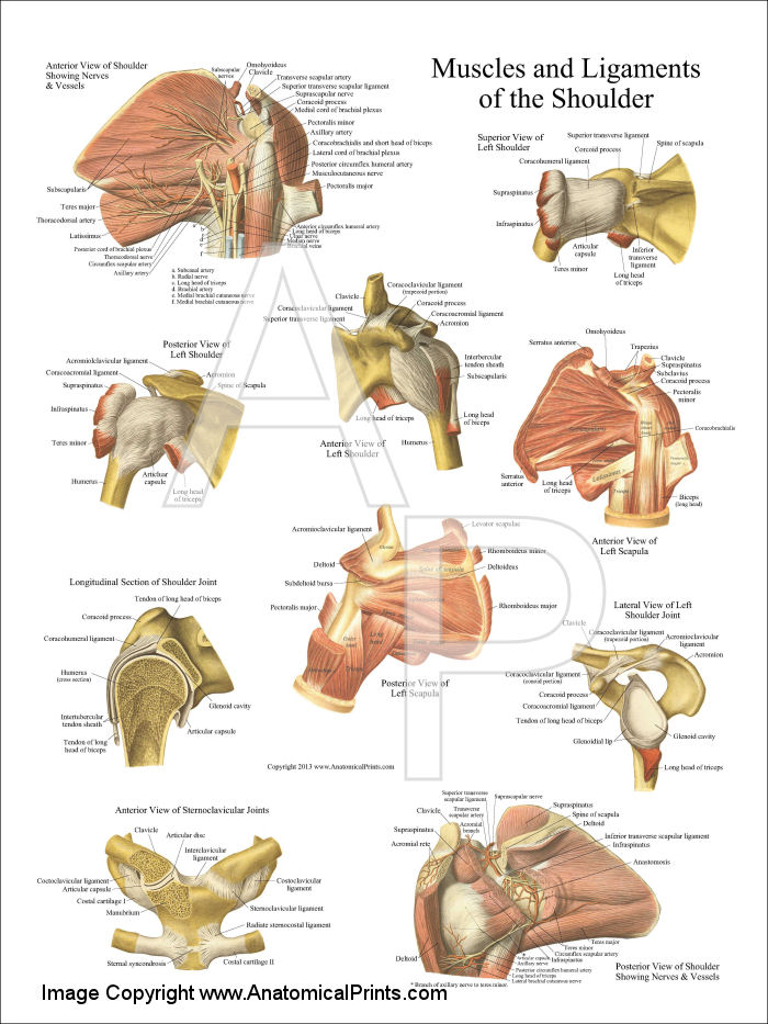
Muscles and Ligaments of the Shoulder Poster from www.dcfirst.com The bicep has two shoulder tendons: The glenoid fossa forms a very shallow socket, so the muscles, ligaments, and cartilage of the shoulder joint reinforce its structure and help to prevent dislocations. Joint diagram unlabeled shoulder ball and socket joint diagram diagram of shoulder joint ligaments. A ring of cartilage known as the labrum surrounds the glenoid fossa to extend the size of the socket while maintaining flexibility. Ligaments, to connect the bones; Atlas of the anatomy of the joint of the shoulder on a ct arthrogram in axial, coronal, and sagittal sections, on a 3d images and on conventional athrogram. The shoulder joint (glenohumeral joint) is a ball and socket joint between the scapula and the humerus. The shoulder isn't just one bone, it's actually made up of three different bones and various tendons, ligaments, and muscles.the three bones located in the shoulder are the humerus, the scapula, and the clavicle.
The shoulder is not a single joint, but a complex arrangement of bones, ligaments, muscles, and tendons that is better called the shoulder girdle.
Shoulder joint diagram — untpikapps from www.untpikapps.com marked elevation of lateral end of clavicle. The joints of the shoulder that are primarily responsible for movement are held together by several strong ligaments. Supraspinatus, infraspinatus, subscapularis, and teres minor. These muscles form the outer shape of the shoulder and underarm. They include the coracoclavicular ligaments, the coracoacromial ligaments, the superior transverse scapular ligament, the coracohumeral ligament, the acromioclavicular ligament, and the glenohumeral ligaments. By ligaments reinforcing the shoulder joint include: The clavicle (collarbone), the scapula (shoulder blade), and the humerus (upper arm bone) as well as associated muscles, ligaments and tendons. This gives the shoulder a wide range of motion, but also makes it vulnerable to injury. Shoulder ligaments can lose strength due to constant movement of the shoulder bones, and muscles. The long head and the short head. A joint capsule is a watertight sac that surrounds a joint. Muscles and ligaments of the shoulder poster. Shoulder complaints were extremely prevalent in my.
Other important bones in the shoulder include: Shoulder tendons chart ~ labeled anatomy chart of shoulder ligaments on white background stocktrek images. Muscle anatomy back 12 photos of the muscle anatomy back back muscle anatomy images, back muscle anatomy of the human body, back pain muscle anatomy, muscle anatomy lower back, posterior back muscle anatomy, human muscles, back muscle anatomy images, back muscle anatomy of the human body, back pain muscle anatomy. The shoulder joint permits a fuller range of motion than any other joint, allowing the arm to raise, lower, extend and rotate a full 360 degrees. Related online courses on physioplus.
Flashcards - Anatomy Block I - Back and Shoulder - Axial ... from classconnection.s3.amazonaws.com The right shoulder, the left shoulder; Diagram of shoulder ligaments : Beyond this, there is also a shoulder joint arrayed in a ball and socket formation, a rotator cuff, and various muscles like the deltoid muscle and the teres major muscle. To ensure proper range of motion, the shoulder joint is supported by the shoulder ligaments, shoulder tendons and shoulder muscles. This gives the shoulder a wide range of motion, but also makes it vulnerable to injury. The long head and the short head. Ac joint is a diathrodial joint with a fibrocartilaginous disk. The fixture included an additional aluminum plate (c) which was connected and moved with the instron actuator.
Fall on one point of shoulder and can rupture these ligaments with dislocation of ac joint.
Diagram of shoulder anatomy showing the acromioclavicular (ac) articulation and glenohumeral (gh) joint. The shoulder has about eight muscles that attach to the scapula, humerus, and clavicle. Shoulder tendons chart ~ labeled anatomy chart of shoulder ligaments on white background stocktrek images. The shoulder is not a single joint, but a complex arrangement of bones, ligaments, muscles, and tendons that is better called the shoulder girdle. Joint diagram unlabeled shoulder ball and socket joint diagram diagram of shoulder joint ligaments. Diagram of the human shoulder joint. The long head and the short head. The shoulder is not a single joint, but a complex arrangement of bones, ligaments, muscles, and tendons that is better called the shoulder girdle. A ligament in the shoulder can rupture due to various reasons. Back muscles diagram and ligaments, human muscles, back muscle diagram, back muscle diagram exercise. Anatomy of the human body via wikimedia commons, public domain. A torn ligament is one such injury that an individual tends to have. These are the coracohumeral, glenohumeral and transverse humeral ligaments.
The shoulder joint is formed where the humerus (upper arm bone) fits into the scapula (shoulder blade), like a ball and socket. The anatomy of the provides the strength and functionality of the upper body. Shoulder tendons chart ~ labeled anatomy chart of shoulder ligaments on white background stocktrek images. A torn ligament is one such injury that an individual tends to have. These are the coracohumeral, glenohumeral and transverse humeral ligaments.
Pin on Exercise from i.pinimg.com The glenoid fossa forms a very shallow socket, so the muscles, ligaments, and cartilage of the shoulder joint reinforce its structure and help to prevent dislocations. Shoulder tendons chart ~ labeled anatomy chart of shoulder ligaments on white background stocktrek images. In the shoulder joint, the ligaments play a key role in stabilising the bony structures. Each of these muscles has its origin on the scapula and inserts around the head of the. The left shoulder and acromioclavicular joints, and the proper ligaments of the scapula. 17 photos of the diagram of shoulder muscles and tendons. A ring of cartilage known as the labrum surrounds the glenoid fossa to extend the size of the socket while maintaining flexibility. A torn ligament is one such injury that an individual tends to have.
The rotator cuff consists of four muscles:
Although three ligaments protect and surround the shoulder joint, most of its stability comes from the powerful muscles and tendons of the rotator cuff. The rotator cuff consists of four muscles: Ac joint is a diathrodial joint with a fibrocartilaginous disk. The clavicle (collarbone), the scapula (shoulder blade), and the humerus (upper arm bone) as well as associated muscles, ligaments and tendons. Several ligaments make up parts of the joint capsule, and these ligaments are important in keeping the shoulder joint in proper position. Supraspinatus, infraspinatus, ters minor,.et), using interactive animations and labeled diagrams. These muscles form the outer shape of the shoulder and underarm. By ligaments reinforcing the shoulder joint include: Tendons, to attach the muscles to the bones. The shoulder isn't just one bone, it's actually made up of three different bones and various tendons, ligaments, and muscles.the three bones located in the shoulder are the humerus, the scapula, and the clavicle. In the shoulder, the joint capsule is formed by a group of ligaments that connect the humerus to the glenoid. 17 photos of the diagram of shoulder muscles and tendons. • coils and patient position:
Supraspinatus, infraspinatus, subscapularis, and teres minor diagram of shoulder. Ligaments are soft tissue structures that connect bones to bones.


0 Komentar